Home / Echocardiography (Echo) Heart Imaging
An echocardiogram also known as Echocardiography or cardiac echo is a simple non-invasive 30-minute ultrasound examination of the heart which reveals both the structure and function of the heart.
This includes both static and dynamic pictures and can determine whether one has had a previous heart attack, an enlarged heart, or any abnormal structural or valvular abnormalities.

An echocardiogram is performed by a highly-skilled cardiac technician sonographer or cardiology specialist and usually takes between 30 and 45 minutes. The patient lies on an exam table, and an ultrasound probe is placed on the chest, and two-dimensional pictures are produced. This cardiac test can indicate heart chamber or valve abnormalities. The findings and the results of the echocardiography test are usually available within two days.
New York Cardiac Diagnostic Center, located in New York, NY, received accreditation by the Intersocietal Accreditation Commission (IAC) in Echocardiography in the areas of Adult Transthoracic and Adult Stress. New York Cardiac Diagnostic Center has undergone an intensive application and review process, followed by a thorough review by a panel of medical experts, and is found to comply with all the standards.
Why is an Echocardiogram performed?
An echocardiogram is performed to evaluate the valves and chambers of the heart in a non-invasive way. The echocardiogram allows doctors to diagnose, evaluate, and monitor:
- Abnormal heart valves;
- Any structural or functional defects of the heart;
- Atrial fibrillation;
- Congenital heart disease;
- Damage to the heart muscle in patients who have had heart attacks;
- Heart murmurs;
- Infection in the sac around the heart (pericarditis);
- Infection on or around the heart valves (infectious endocarditis);
- Pulmonary hypertension;
- The heart’s pumping ability;
- The pumping function of the heart for people with heart failure;
- The source of a blood clot after a stroke or TIA;
- The structure, thickness and movement of the heart valves;
- The size of the chambers of the heart, which can change with conditions such as hypertension, heart damage with a myocardial infarction or congestive heart failure.
If you experience any cardiovascular signs or symptoms, visit our cardiology office in nearby New York City locations, including Downtown, Midtown, and Upper East Side, and meet Dr. Steven Reisman. He is an expert specialist who can provide patients with echocardiography testing and analysis.
I have known Dr. Reisman for many years. My first experience with him was through a friend’s referral. A family situation came along where we needed a cardiologist, a great one. I am very thankful that I was referred to Dr. Reisman. He is a true leader in his field; very caring for his patients and he will go out of his way to help anyone. Dr. Reisman is very approachable and friendly. His office staffs are very friendly. After attending to my family situation I too was in need of a cardiologist for stress testing – I have been to the Wall Street & Upper East Side location – one close to work and one close to home. Again, my experience was 100% positive. If you are looking for a cardiologist for echo or stress testing, don’t look any further.
Elliot
Dr. Reisman is professional and well knowledge in his field . He only orders exams that are required. The techs are also professional, and make you feel comfortable during the procedures. Dr. Reisman office manager Michelle, is pleasant and explains the billing process, before any tests are performed. Dr. Reisman thank you for want you do.
AD
Dr. Reisman is an amazing cardiologist with a true heart of gold. I was so impressed with his thoroughness and kindness. He truly helped me and was never rushed. He really cares. Highly recommended. He really is the best of the best. Very knowledgeable, experienced, empathetic, caring and professional. He always makes sure to address all concerns and make them feel comfortable so they can trust him. Also very well mannered and engaging!
Erica B.
When to see a doctor?
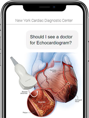
 You should call a specialist who specializes in echocardiography if you experience symptoms of heart disease, like shortness of breath, chest discomfort, or swelling in the legs, or if you have an existing condition such as a heart murmur. This test is performed in the doctor’s office by a trained sonographer or Dr. Reisman.
The appointment for the Echocardiogram procedure takes about 40 minutes. The results are usually available within several days.
What can the results of the Echocardiogram show?
The result can help determine if you are at risk for a heart attack or heart failure. It can also indicate if further testing is necessary or if you need a treatment plan.
- The size and shape of your heart;
- How well your heart is working overall (that is how well it contracts and how well it relaxes);
- If a wall or section of heart muscle is weak and not working correctly;
- If you have problems with your heart’s valves;
- If you have a blood clot or fluid around your heart.
What Can an Echocardiogram Detect?
 Echocardiograms, which show the structure and function of the heart, are used to diagnose the following conditions:
- Regurgitation (mitral valve of the heart does not close tightly, allowing blood to flow backward in the heart)
- Stenosis (heart’s aortic valve narrows, blocks blood flow from your heart into the main artery)
- Pericarditis (swelling and irritation of the sac surrounding the heart)
- Congestive heart failure (heart doesn’t pump blood as efficiently as it should)
- Aneurysms (enlargement of an artery caused by weakness in the arterial wall)
- Blood clots (blood clots form in the blood vessels and obstruct blood flow, causing blockages that affect the heart and lungs)
- Cardiomyopathy (heart enlargement or thickening that makes it difficult for the heart to deliver blood to the body and can lead to heart failure)
- Atrial septal defect (abnormal opening between the heart’s upper chambers
- Valvular heart disease (narrowing, leaking or slipping out of place of heart valves)
- Atherosclerosis (fatty material, cholesterol, and other substances accumulate on the inner artery walls)
What to Expect
An echocardiogram necessitates some preparation. There are also pre-and post-procedural steps to take. Read on to find out more about what to expect.
Before the Procedure
The doctor will explain the procedure and allow you to ask questions. There is no need for any prior preparation, such as fasting or sedation.
- Inform your doctor about all medications (both prescription and over-the-counter) and herbal supplements you are taking.
- Inform your doctor if you have any devices in your chest that help you control your heartbeat.
During the Procedure
An echocardiogram test can be done as an outpatient procedure or as part of a hospital stay. Procedures may differ depending on your condition and the practices of your doctor.
This is usually followed by an echocardiogram:
- You will be asked to take off any jewelry or other objects that could obstruct the procedure. You may be required to wear glasses, dentures, or hearing aids.
- You will be asked to remove your clothes and given a gown to wear.
- You will be lying on your left side on a table or bed. For added support, place a pillow or wedge behind your back.
- You’ll be connected directly to an ECG monitor, which records the electrical activity of your heart and monitors it during the procedure with small, adhesive electrodes.
- The room will be darkened so that the technologist can see the images on the echo monitor.
- The technologist will apply warmed gel to the patient’s chest before placing the transducer probe on the gel.
- The technologist will move the transducer probe around and apply varying amounts of pressure during the test to obtain images of various locations and structures of the heart. The amount of pressure applied behind the probe should not be painful.
After the Procedure
Unless your doctor advises otherwise, you may resume your regular diet and activities. In general, no special care is required following an echocardiogram. However, depending on your needs, your doctor may provide additional or alternate instructions following the procedure.
How Exactly Does An Echocardiogram Work?
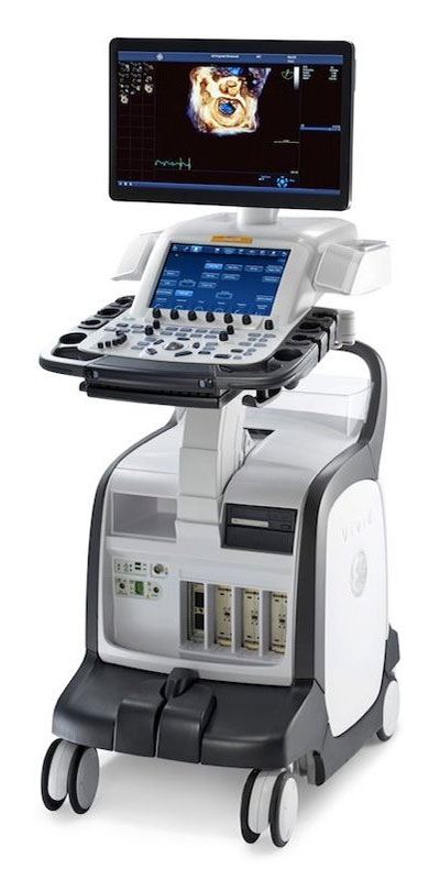
Sound waves are sent by the transducer towards the heart. Sound waves bounce back like an echo, similar to sonar from a submarine. The echoes of the heart are then gathered by the transducer. The echoes are processed on a computer and a 2D image is produced on a screen. Through the transducer, the echocardiogram allows vital areas of the heart to be examined.
What Is the Difference Between a Transthoracic Echocardiogram (Tte) And a Transesophageal Echocardiogram (TEE)?
A transthoracic echocardiogram (TTE) is the standard echocardiogram usually done in a physician’s office or a hospital setting. An ultrasound technologist places gel on the patient’s chest and then applies a transducer to capture the heart’s ultrasound images. The patient for this examination needs no special preparation.
A transesophageal echocardiogram (TEE) is performed when one cannot get adequate images of a standard echocardiogram. This examination is more invasive, and a tube with a transducer is placed down the patient’s mouth and into the esophagus to capture pictures of the heart.
The transesophageal echocardiogram (TEE) procedure frequently requires sedation and has special preparation, including not eating or drinking for 6 hours before the test, and is usually done in a hospital setting.
Echocardiogram vs EKG
An electrocardiogram is also known as an EKG or ECG is a measurement on a graph of the electrical activity of the heart. This measurement is performed by placing little patches called electrodes on the patient’s chest in various positions and then attaching them with lead wires to a machine that records the electrical activity of the heart. This test can be useful in detecting irregular heartbeats, enlargement of the heart, electrical and conduction abnormalities, and prior heart attack. The electrocardiogram has limited diagnostic capabilities.
An echocardiogram is also known as an ultrasound of the heart provides a greater degree of information by utilizing dynamic pictures of the heart. The echocardiogram is done by placing gel on the patient’s chest and using a transducer to relay images of the heart function and structure in a very comprehensive examination. The echocardiogram can be useful at both rests and before and after exercise in evaluating heart size, prior heart attack, risk of future heart attack, heart enlargement, and leaky valves in the heart. While an EKG may be performed in only 5 to 10 minutes an echocardiogram usually requires 20 to 30 minutes.
Finding Echocardiogram Near Me
When looking for an effective first-line diagnostic test to evaluate cardiac disease and function, choose a Cardiac Diagnostic Center stuffed with a board-certified imaging cardiologist as well as an echocardiography technician and a trained sonographer that offer a non-invasive echocardiogram near you. Dr. Steven Reisman offers the latest in imaging techniques and diagnostic heart tests, such as an echocardiogram, providing an effective imaging modality for assessing cardiac structure, function, and hemodynamics to diagnose cardiomyopathy, valvular heart disease, aorta, and pericardial disease.
If you have any questions or would like to schedule a consultation please feel free to contact a leading echocardiogram specialist Dr. Steven Reisman of the New York Cardiac Diagnostic Center and indicate which office (Upper East Side, Midtown Manhattan, or Wall Street / Financial District) you would like to see the cardiologist for the echocardiogram in NYC.

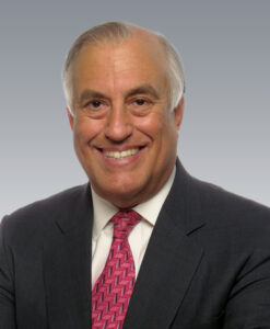
Dr. Steven Reisman is an internationally recognized cardiologist and heart specialist. He is a member of the American College of Cardiology, American Heart Association, and a founding member of the American Society of Nuclear Cardiology.
Dr. Reisman has presented original research findings for the early detection of "high risk" heart disease and severe coronary artery disease at the annual meetings of both the American College of Cardiology and the American Heart Association. Dr. Reisman was part of a group of doctors with the Food and Drug Administration who evaluated the dipyridamole thallium testing technique before the FDA approved it.
Dr. Steven Reisman's academic appointments include Assistant Professor of Medicine at the University of California and Assistant Professor at SUNY. Hospital appointments include the Director of Nuclear Cardiology at the Long Island College Hospital.
